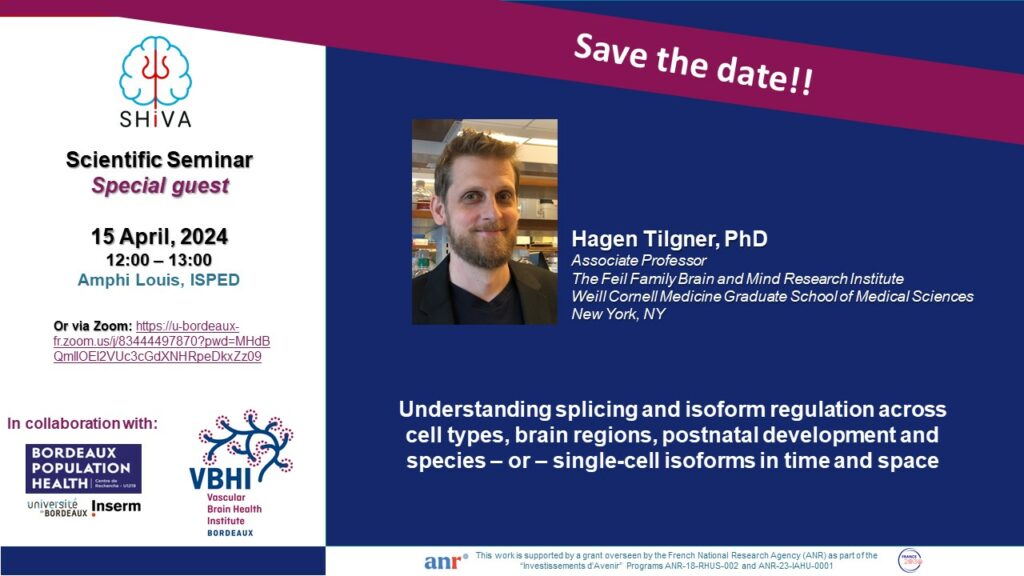
Hagen Tilgner, PhD, studied computer science in Germany and France, and after a brief stint at the Sanger Institute (UK), did his PhD with Roderic Guigó at the Centre for Genomic Regulation in Barcelona. There he focused on RNA and the co-transcriptionality of splicing (see Tilgner et al, Genome Res, 2012 for example). His postdoctoral work at Stanford with Michael Snyder focused on technology development, specifically for long-read transcriptomics (see for example Sharon, Tilgner, Grubert, Snyder, Nature Biotechnology, Tilgner, Sharon, Grubert, Snyder, PNAS or Tilgner, Jahanbani* et al, Nature Biotechnology). He started his lab at Weill Cornell in New York City in 2016 focusing on technologies to decipher the actions of RNA isoforms in the brain. The lab is a multi-disciplinary lab, including wet-lab technology development (see for example single-cell isoform RNA sequencing, ScISOr-Seq, Gupta et al, Nature Biotechnology or SnISOr-Seq, Hardwick et al, Nature Biotechnology) and dry-lab approaches (see for example Joglekar et al, Nature Communications or Prjibelski et al, Nature Biotechnology) as well as combined large-scale efforts centered on the brain (for example, Joglekar et al, Nature Neuroscience, in press) where Maths/CS, molecular biology and neuroscience backgrounds interact to further our understanding of isoforms in healthy and diseased brain of humans and model organisms.
Most mammalian genes encode multiple distinct RNA isoforms and the brain harbors especially diverse isoforms. Complex tissue includes diverse cell types, which employ distinct isoforms. To untangle cell-type specific brain isoform profiles, we developed the first single-cell long-read technology for >>1,000 cells and fresh tissues (Single-cell isoform RNA sequencing – ScISOr-Seq)1 as well as for frozen tissues (Single-nuclei isoform RNA sequencing – SnISOr-Seq2). To add spatial resolution, we developed Slide-isoform sequencing (Sl-ISO-Seq)3. Collectively, these long-read approaches reveal a striking difference between coordinated pairs of exons with in-between exons (“Distant coordinated exons”) and without in-between exons (“Adjacent coordinated exons”): The former show strong enrichment for cell-type specific usage of exons, whereas the latter do not in mouse1 and human brain2. Of note, coordinated TSS-exon pairs and exon-polyA-site pairs follow the same trend as distant coordinated exon pairs2. Simultaneously, autism-associated exons are among the most highly variably used exons across cell types2. Spatially barcoded isoform sequencing revealed that often region-specific isoform differences correlate with precise boundaries of brain structures (e.g., from the choroid plexus to the hippocampus). However, genes including Snap25 go against this trend, using a steady gradient of exon inclusion as one traverses the brain3. Moreover, choroid plexus epithelial cells show a dramatically distinct isoform profile, which originates from distinct exon and poly(A) site usage, but most strongly from distinct TSS usage3. For the NIH Brain Initiative, we have mapped single-cell isoform expression across development, brain regions and species. Neurotransmitter release and reuptake as well as synapse turnover genes harbor variability in the same cell type across anatomical regions but the same cell type traced across development shows more isoform variability than across adult anatomical regions. Moreover, most cell-type specific exons in adult mouse hippocampus behave similarly in human hippocampi. However, human brains have evolved additional cell-type specificity in splicing, suggesting gain-of-function isoforms4. Most recently, we have made advances in understanding the error sources of Pacific Biosciences and Oxford Nanopore long-read sequencing technologies5 and have implemented highly accurate long-read interpretation software6. Finally, I will comment on further, yet unpublished, technologies of the lab.
1.Gupta, Collier et al, Nature Biotechnology, 2018; 2.Hardwick, Hu, Joglekar* et al, Nature Biotechnology, 2022; 3.Joglekar et al, Nature Communications, 2021; 4. Joglekar et al, Nature Neuroscience, in press; 5. Mikheenko, Prjibelski et al, Genome Research, 2022 ; 6. Prjibelski, Mikheenko et al, Nature Biotechnology, 2023